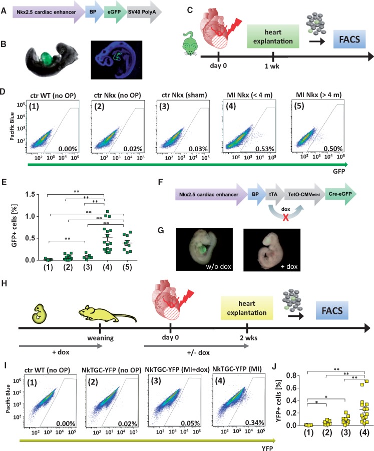
Figure 1.
Identification of NkxCE-positive cells after myocardial infarction (MICs). Vector construct of the Nkx2.5 cardiac enhancer GFP (NkxCE-GFP) mouse (A). (B) Cardiac-specific GFP-fluorescence in an NkxCE-GFP embryo (E9.5). Left picture shows whole-mount in situ GFP-fluorescence, whereas the right picture shows a cryo-section stained for GFP (green) (blue: DAPI). (C) MI was induced by ligation of the LAD in adult NkxCE-GFP mice; heart explantation after 1 week. Single-cell suspension was cardiomyocyte-depleted via several filter steps. Quantification of GFP+ cells by FACS. (D) Amount of GFP+ MICs detected by FACS (exemplary plots). (1) Ctr WT (no OP): Wild type mouse (C57Bl/6) without surgery. (2) Ctr Nkx (no OP): NkxCE-GFP mouse without surgery. (3) Ctr Nkx (sham): NkxCE-GFP mouse with sham operation. (4) MI Nkx (<4 m): NkxCE-GFP mouse with MI (< 4 months). (5) MI Nkx (>4 m): NkxCE-GFP mouse with MI (>4 months). (E) About 0.5% GFP+ MICs were detected in mice with MI but not in the control groups. [groups see D., n(1) = 19, n(2) = 15, n(3) = 6, n(4) = 16, n(5) = 9]. **P < 0.01 (Kruskal–Wallis, postHoc: Dunn’s method). (F) Vector construct of the transgenic NkxCE-tTA-Cre-GFP (NkTGC) mouse. (G) Left panel shows cardiac-specific whole-mount in situ YFP-fluorescence in an NkTGC x Rosa26YFP embryo (E9.5) (without doxycycline substitution in the dam’s drinking water). Right panel shows no YFP-fluorescence in an E9.5-embryo substituted with dox (+ dox) in the dam’s drinking water during embryonic development. (H) MI was induced by LAD-ligation in adult NkTGC x Rosa26YFP (NkTGC-YFP) mice substituted with dox from embryonic development until weaning indicating no YFP-fluorescence in their hearts. As a control, half of the MI-surgery group received dox-enriched drinking water from 1 week before LAD-ligation to monitor the ‘leakiness’ of the system; heart explantation 14 days after MI. Quantification of YFP+ cells by FACS. (I) Amount of YFP+ cells detected by FACS (exemplary plots). Groups: (1) Ctr WT (no OP): Wild type mouse (C57Bl/6) without surgery. (2) Ctr NkTGC-YFP (no OP): NkTGC-YFP mouse without surgery. (3) Ctr NkTGC-YFP (MI+Dox): NkTGC-YFP mouse with MI (achieved dox by drinking water from 1 week before MI until heart explantation). (4) NkTGC-YFP (MI): NkTGC-YFP mouse with MI. (J) About 0.25% of YFP+ cells were detected in mice with MI (group 4) but not in the control groups [groups see I., n(1) = 5, n(2) = 7, n(3) = 10, n(4) = 15]. *P < 0.05; **P < 0.01 (Kruskal–Wallis, postHoc: Dunn’s method). FACS, fluorescence-activated cell sorting.

