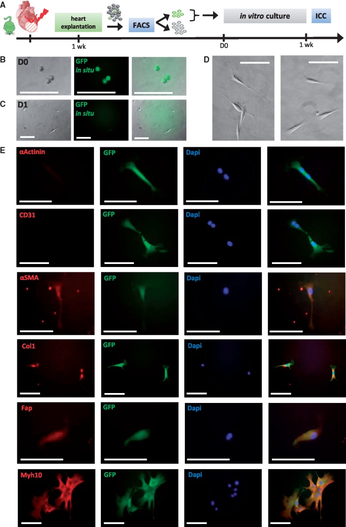
Figure 6.
In vitro culture of NkxCE-GFP+ MICs isolated after MI. (A) Hearts from infarcted NkxCE-GFP mice were explanted 1 week after MI (n = 6). 20 GFP+ MICs were directly sorted to collagen-coated 96-well-plates. Immunocytochemical staining was performed after 1 week. (B) Directly after sorting (D0) GFP+ MICs exhibit a round shape and in situ GFP fluorescence (no antibody). Scale bars: 100 µm. (C) One day later (D1), in situ GFP-fluorescence has faded and 3 days after sorting (D), GFP-fluorescence was no longer visible. Morphologically, cells appeared fibroblast-like. Scale bars: 100µm. (E) Immunocytochemical staining 1 week after plating. αActinin and CD31 were not detected. However, fibroblast/myofibroblast markers like αSMA, Col1, Fap, and Myh10 were detected in in vitro cultivated MICs. Scale bars: 100 µm

