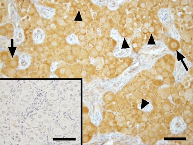
Figure 2.

Photomicrographs of VASA expression in ovarian tissue of a representative fetal macaque under ×400 magnification. Scale bar = 50 μm and 65 μm (inserts). Arrow = newly formed primordial follicles. Arrowheads = germ cells still in germ cell nest. Primary antibody omission negative control (inserts) showed a lack of nonspecific binding.
