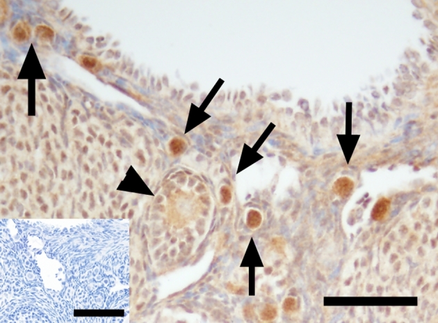
Figure 4.

FOXO3 immunoreactivity (brown, arrows) in B6;129 mouse ovarian tissue in a ×400 magnification image. Arrows indicate positive FOXO3 staining in the nucleus of primordial oocytes. Arrowheads show cytoplasmic staining of FOXO3 in the mouse primordial oocyte. Primary antibody omission negative control (insert) showed a lack of nonspecific binding. Scale bar = 50 μm and 65 μm (insert).
