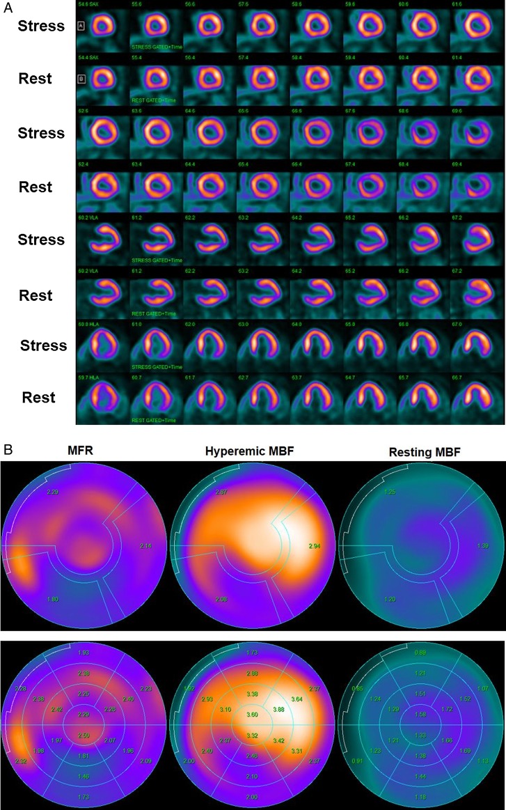
Figure 1.
Normal myocardial perfusion and MBF study with 13N-ammonia PET/CT in a 45-year-old women with atypical chest pain and borderline arterial hypertension. (A) Regadenoson-stress and rest 13N-ammonia PET/CT images in corresponding short-axis (top), vertical long-axis (middle), and horizontal long-axis (bottom) slices. As can be appreciated, there is normal and homogeneous radiotracer uptake of the left ventricular wall on stress and rest 13N-ammonia PET/CT, indicative of normal perfusion. (B) Regional MBF quantification signifies normal hyperaemic MBFs and MFR in all three major coronary artery territories of the LAD, LCx, and RCA with hyperaemic MBFs ≥1.8 mL/g/min (upper-middle panel). Calculated MFR is widely normal (≥2.0) except for a borderline reduction in the RCA distribution (upper-right panel). Notably, there is no decrease but rather a mild increase in segmental MBF from the mid- to distal segments and, thus, no abnormal MBF gradient during hyperaemic MBFs is noted (lower-middle panel).

