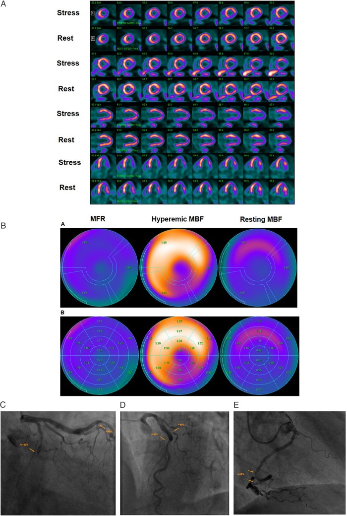
Figure 3.
Abnormal stress-rest myocardial perfusion and MBF study with 13N-ammonia PET/CT in a 64-year-old man with atypical chest pain and previous percutaneous intervention of a LAD lesions. (A) Regadenoson-stress and rest 13N-ammonia PET/CT images in corresponding short-axis (top), vertical long-axis (middle), and horizontal long-axis (bottom) slices. On rest images, there is a mild decrease of myocardial perfusion of the inferior and inferolateral wall to suggest mild necrosis that, however, markedly worsens during vasomotor stress signifying large size and severe ischaemia in the LCx distribution. (B) Regional MBF quantification demonstrates abnormally reduced hyperaemic MBFs and MFR in all three major coronary artery territories of the LAD, LCx, and RCA, respectively (upper panel). Segment MBF analysis outlines a decrease in MBF from the mid- to distal segments with a mean longitudinal MBF gradient during hyperaemic flow in the LAD (0.18 mL/g/min), LCx (0.56 mL/G/min), and RCA (0.09 mL/g/min) (lower-middle panel). (C) Invasive coronary angiography of the left coronary artery in this patient demonstrated a complete occlusion of the mid LCx, which is responsible for the stress-induced ischaemia in the inferior and inferolateral wall, and a ≈50% stenosis in the mid-LAD just after a patent stent (left panel). (D) An additional left-anterior-oblique projection, however, unmasks a ≈95% stenosis of the proximal diagonal branch in addition to the described LAD lesion (right panel). Invasively measured FFR of the LAD lesions proved to be normal with 0.84. (E) Invasive coronary angiography of the right coronary artery demonstrates two serial lesions of ≈50% in the mid- and distal RCA, respectively.

