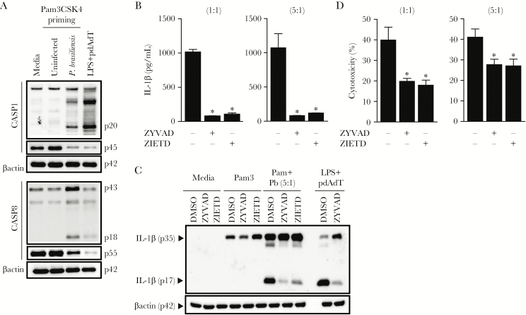
Figure 2.
Caspase-8 signaling is important for Paracoccidioides brasiliensis-triggered interleukin (IL)-1β processing and release. (A) wild-type (WT) bone marrow-derived dendritic cells (BMDCs) were primed with 200 ng/mL Pam3CSK4 for 3 hours and infected with P brasiliensis (multiplicity of infection 5) for 24 hours before lysates, and supernatants were collected and immunoblotted for caspase-8 and caspase-1, respectively. Control cells were stimulated with canonical inflammasome activator pdAdT after lipopolysaccharide priming. (B) Culture supernatants harvested from WT BMDCs activated for 3 hours with Pam3CSK4, treated the last 2 hours with caspase-1 (z-YVAD-fmk) or caspase-8 (z-IETD-fmk) inhibitors, and subsequently challenged for 24 hours with P brasiliensis were analyzed either by enzyme-linked immunosorbent assay to measure the levels of secreted IL-1β, or (C) a combination of supernatants and cell lysate were separated by sodium dodecyl sulfate polyacrylamide gel electrophoresis, blotted, and probed with a monoclonal IL-1β antibody. (D) Pam3CSK4-primed BMDCs untreated or treated with z-YVAD-fmk or z-IETD-fmk were cultured with P brasiliensis before the assessment of extracellular lactate dehydrogenase. Data show the averages ± standard deviation from triplicate wells of at least 3 independent experiments. The symbol (*) indicates a significant difference between dimethyl sulfoxide (DMSO)-treated samples (B and D) (P < .05).

