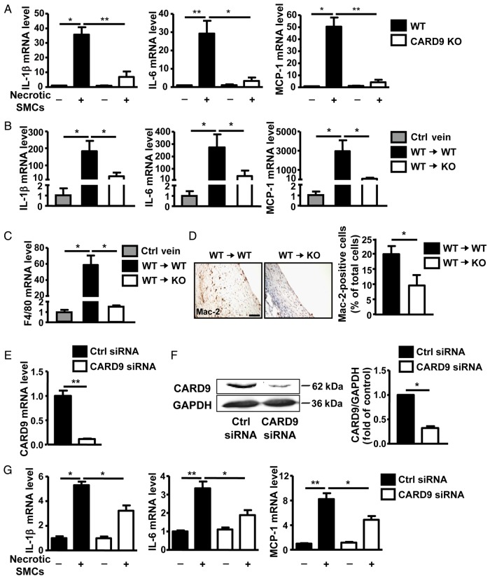
Figure 4.
CARD9-KO attenuated the level of the necrotic SMC-induced pro-inflammatory cytokines in vitro and in vivo. (A) BMDMs derived from WT or CARD9-KO mice were stimulated with necrotic SMCs (H2O2 treated for 8 h) for 20 h. qPCR analysis of the expression of IL-1β, IL-6, and MCP-1 expressed as fold induction compared with the control (Ctrl) after normalization to α-tubulin. Data represent the mean ± SEM; five independent experiments were performed. The mRNA expression of (B) IL-1β, IL-6, MCP-1, and (C) F4/80 in control veins and vein grafts at 3 days presented as fold induction compared with the control after normalization to α-tubulin; n = 6 per group. (D) Expression of Mac2 in sections of vein grafts at 4 weeks from WT and CARD9-KO mice and quantification. Data represent the mean ± SEM; n = 6 per group. THP1 cells were transfected with CARD9 or control siRNA for 36 h; (E) mRNA and (F) protein expression of CARD9 show as fold change compared with the control siRNA group after normalization to α-tubulin and GAPDH, respectively. (G) THP1 cells with or without CARD9 knockdown were stimulated with necrotic HSVSMCs (H2O2 treated for 8 h). qPCR analysis of the expression of IL-1β, IL-6, and MCP-1 expressed as fold induction compared with the control after normalization to α-tubulin. Data represent the mean ± SEM; five independent experiments were performed for (E) and (G), and three independent experiments were performed for (F). Scale bar: 50 µm. *P < 0.05, **P < 0.01.

