Figure 2.

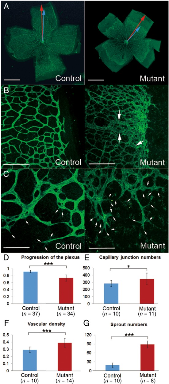
Abnormal development of retinal vasculature in Tmem100-iKO newborn mice. (A) FITC-conjugated isolectin B4 labelling showed complete formation of superficial vascular plexus in the control mice but delayed progression of the angiogenic front in the Tmem100-iKO mutant mice. The progression of the vascular plexus towards the edge of the retina was measured by the ratio of the vascular radius (blue arrow) and the retina's radius (red arrow). (B) Dilated and hyperbranched vascular plexus were observed in the mutants. Note that these defects were severer in the angiogenic front region. Arrows indicate abnormally dilated vessels resembling AV shunts. (C) High magnified image of the angiogenic front region represented increased number of filopodial protrusions (arrows) in the tip cells. (D–G), Comparison of several angiogenic parameters between the control and mutant retinas; progression of the vascular plexus towards the edge of the retina (D), number of capillary junctions within a field of view (×200) (E), vascular density (the covered area by blood vessels in the vascular plexus region) within a field of view (×25) (F), number of sprouts (filopodial protrusions in the tip cells) within a field of view (×100) (G). *P < 0.05, ***P < 0.001. Scale bars: A, 1 mm; B, 200 μm; C, 100 μm.
