
Figure 3.
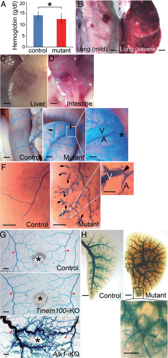
Vascular defects of Tmem100-iKO adult mice. (A–D) Reduced haemoglobin level (Control, n = 11; Mutant, n = 11, *P < 0.05) (A) and signs of internal haemorrhages in the lungs (B: left panel, mild haemorrhage; right panel, severe haemorrhage; 8/16), liver (C; 6/6), and intestine (D; 8/16) of Tmem100-iKO mutants. Two-tailed Student's t-test was performed for statistical analysis. (E–H) Blood vessels in the small intestine (E), liver (F), back skin (G), and lung (H) visualized by latex dye perfusion. Presence of the latex dye in both arteries (A) and veins (V) would indicate AV shunts (arrows). (E) The focal AV shunts in the intestine were detected close to haemorrhagic areas indicated by an asterisk. Control, n = 7; Mutant, n = 5 (four of them showed AV shunts). (F) Note direct connection (arrowhead) between arteries (A) and veins (V) in the AV shunts of the liver. Control, n = 7; Mutant, n = 8 (four of them showed AV shunts). (G) No sign of AV shunts was observed in the wounded skin area of control (n = 4) and Tmem100-iKOs (n = 4), compared with wounded skin of tamoxifen-treated Alk12loxP/2loxP;ROSA26+/CreER mice (Alk1-iKO; n = 20). *, wound; red arrowheads, arteries; blue arrowheads, veins. (H) Leakage of the latex dye was detected in the pulmonary vasculature. Scale bars: B–H, 1 mm; enlarged images, 0.25 mm.