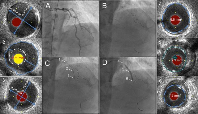
Figure 1.
Ultra-low contrast coronary angiography followed by staged percutaneous coronary intervention with zero contrast. Cine images recorded at the initial angiography using ultra-low contrast volume are displayed on adjoining screen during the staged percutaneous coronary intervention (A) and used to guide catheter engagement, coronary guide wire placement in the left anterior descending artery, diagonal branch, and the circumflex artery, thus creating a metallic silhouette of the left coronary system (B). Intravascular ultrasound imaging of the left anterior descending artery is performed with proximal reference diameter (≈4.5 mm) (1), minimal luminal area (3.71 mm2) (2), and distal reference diameter (≈4.0 mm) (3) measured for selection of the appropriate pre-dilation balloon and stent sizes. The co-registered dry cine image of intravascular ultrasound transducer placed at the distal reference (C) is used to guide the percutaneous coronary intervention. Following preparation of the lesion and deployment of a 3.5 × 38 mm drug-eluting stent (D), intravascular ultrasound is repeated to assess the result, to determine the proximal (9.6 mm2) (4) and distal (7.7 mm2) (6) reference areas, and to guide post-dilation of under-expanded segments to achieve the pre-determined MSA, defined as >90% of the mean of the proximal and distal reference areas, (7.9 mm2) (5).

