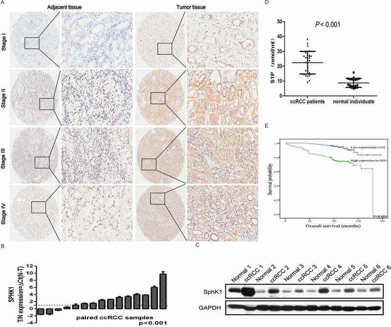
Figure 1.

SphK1 expression is upregurated in ccRCC.
Representative immunohistochemistry micrographs of SphK1 expression from the TMAs among 290 ccRCC patients (left 5 × magnification, right 200 × magnification). The expression of SphK1 was observed within the cytoplasm of cancer cells with weak staining in the stage I localized ccRCCs, weak to intermediate staining in II and III ccRCCs, and strong staining in stage IV metastatic ccRCCs. B. Quantitative RT-PCR assay for SphK1 mRNA level in 15 pairs of human clinical ccRCC tumor tissue and adjacent normal tissue. SphK1 mRNA expression levels were normalized to GAPDH mRNA expression. C. Western blot analysis of SphK1 protein in primary ccRCC tumor tissues and adjacent normal tissues. Expression levels were normalized with GAPDH. D. LC-MSMS analysis for S1P levels in plasma from a small group of ccRCC patients (N = 30) and normal individuals (N = 20). Average S1P concentration was 22.36 ± 7.62 nmol/ml for ccRCC patients versus 8.78 ± 3.04 nmol/ml for normal control with a significant difference (P < 0.001). E. Kaplan-Meier survival analysis revealed that patients with high SphK1 expression had significantly lower 5-year overall survival rates, P < 0.001.
