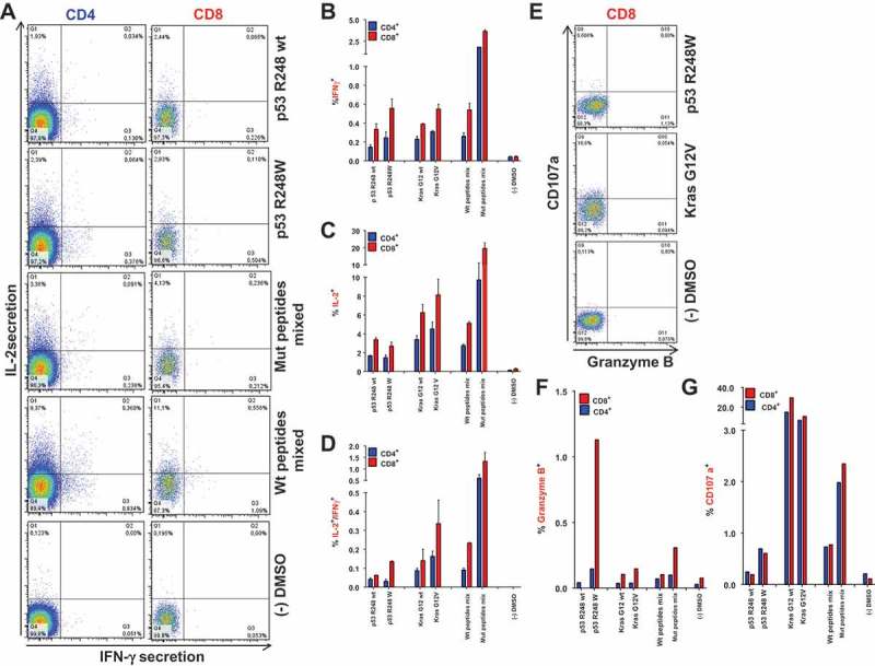
Figure 3.

Vaccination-induced, mutation-specific T cells show poly-functionality. (A) IFNγ/IL-2 two-color cytokine secretion assay with splenic T cells purified from A2.DR1 dtg mice immunized with the most-immunogenic peptides (p53 R24Q/W, Kras G12V/D/R, vaccine regimen: peptides in PBS-based formulations including 50 μg CpG ODN 1668 as an adjuvant, twice on a bi-weekly basis). Cell surface markers were stained with fluorescently labeled monoclonal antibodies against IFN-γ (detection Ab), IL-2 (detection Ab), CD4, CD8, CD11c and NK1.1 (samples shown: T cells re-stimulated on DCs pulsed with wt peptide p53 R248 wt, mutated peptide p53 R248W, most immunogenic mutated peptides mixed; negative control: dendritic cells pulsed with peptide solvent (DMSO) only) . (B)-(D) Quantification of IFNγ/IL-2 two-color cytokine secretion assay shown in (A). In vitro recall responses were obtained from two-color cytokine secretion assays (IL-2, IFN-γ) with pan-T cells purified from immunized A2.DR1 dtg mice. Percentages of IFN-γ (B), IL-2 (C) and IFN-γ/IL-2 (D) double-positive of CD8+ and CD4+ T cells upon in vitro recall against single peptides, corresponding wt peptides and against whole peptide mixes (mix) presented by CD11c+ DCs are displayed. Results are plotted as means of triplicate assays ± SEM. (E) CD8+ effector T cells derived from Kras G12V and p53 R248W mutated peptides vaccinated mice express markers of cytotoxicity upon in vitro antigen-specific re-stimulation. Effector pan-T cells derived from A2.DR1 dtg mice vaccinated with Kras G12V/p53 R248W mutated peptides were in vitro re-stimulated for 12 h on different peptide pulsed DCs. Fluorescently-labeled anti-CD107a mAb and GolgiStop reagent were added to the co-culture. Thereafter, live-dead staining was performed and samples were fixed. Fixed cells were stained for markers granzyme B, IFN-γ, CD11c, CD4, and CD8 with fluorescent-labeled mAbs. CD107a plotted against granzyme B in CD8+ T cells are shown. Samples displayed: T cell re-stimulated on DCs pulsed with mutated peptide p53 R248W and mutated peptide Kras G12V, negative control: DCs pulsed with peptide solvent (DMSO) only. Percentages of granzyme B (F) and CD107a (G) positive of CD8+ and CD4+ T cells from all different peptide samples tested are shown.
