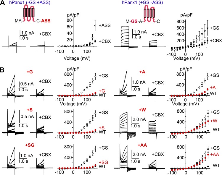
Figure 2.
N-terminal insertion alters voltage-dependent Panx1 channel activity. (A) Whole-cell recordings of HEK cells expressing hPanx1+ASS (left) or hPanx1+GS (right). Cells were held at −60 mV and stepped between −100 and +160 mV for 1.0 s in 20-mV increments. CBX (50 µM) was applied using a rapid solution exchange system. Shown are representative recordings from at least three different cells. Each point represents the mean current density at each voltage, and bars represent SEM. (B) Whole-cell recordings of hPanx1 constructs featuring variable amino acid insertions immediately following the start methionine. Recordings were obtained from transfected HEK cells held at −60 mV and stepped between −100 and +160 mV for 1.0 s. Each point represents the mean current density at each voltage, and bars represent SEM.

