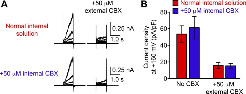
Figure 4.
CBX inhibits Panx1 only from the extracellular side. (A) Representative recordings from WT hPanx1–transfected HEK cells with normal internal solution (top) or internal solution containing 50 µM CBX (bottom). Cells were held at −60 mV and equilibrated for ∼3 min before stepping between −100 and +160 mV for 1.0 s in 20-mV increments. External CBX (50 µM) was applied using a rapid solution exchanger. Shown are representative recordings for four different cells. (B) Current density of recordings shown in A. Bars represent the mean current density of patches obtained with either internal solution with and without external CBX application, and error bars represent SEM. Student’s t test revealed no statistically significant difference with and without internal CBX.

