Figure 1.
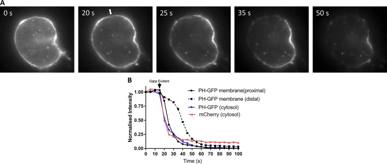
Digitonin permeabilization reveals reversible binding of PH-GFP to the plasma membrane of chromaffin cells. Bovine chromaffin cells were cotransfected with plasmids encoding PH-GFP and mCherry and imaged 5 d later in epifluorescence. Cells were bathed in Na glutamate solution (139 mM Na glutamate, 20 mM PIPES, 0.5 mM EGTA, 0.5 mM MgCl2, and 2 mM ATP) at 27°C and individually perfused with bath solution containing 10 µM digitonin through a 100-µm-inner-diameter glass pipette. (A) Within 30 s of digitonin application, gaps appeared in the PH-GFP–labeled plasma membrane (arrow) and a wave of loss of membrane PH-GFP fluorescence occurred starting at the plasma membrane proximal to the gaps. (B) PH-GFP intensities of segments of the plasma membrane proximal and distal to the gaps and in the cytosol were measured. Cytosolic mCherry fluorescence is rapidly lost coincident with the appearance of a gap in the plasma membrane, and PH-GFP fluorescence on the plasma membrane proximal to the gap decreases almost as rapidly as cytosolic mCherry and cytosolic PH-GFP. These results are similar to those from three other cells in which an initial gap in the PH-GFP fluorescence was detected.

