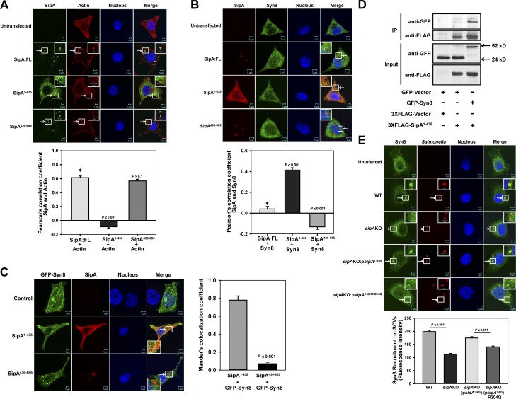
Figure 3.
N-terminal domain of SipA recruits Syn8 on SCV. (A) SipA:FL, SipA1–435, or SipA436–685 were expressed in HeLa cells as N-terminal FLAG-tagged proteins, and cells were stained with phalloidin (red) and anti-FLAG antibody (green). (B) SipA:FL, SipA1–435, or SipA436–685 was expressed in HeLa cells as above, and cells were stained with anti-FLAG antibody (red) and anti-Syn8 antibody (green). (C) HeLa cells were cotransfected with GFP-Syn8 (green) and FLAG-tagged SipA1–435 or SipA436–685 as described in Materials and methods. Cells were stained with anti-FLAG antibody (red). Colocalization of SipA1–435 and GFP-Syn8 was determined by confocal microscopy. (D) Immunoprecipitation (IP) of GFP-Syn8 from FLAG-SipA1–435 and GFP-Syn8 coexpressing HeLa cell lysate using immobilized anti-FLAG antibody. Binding of GFP-Syn8 with FLAG-SipA1–435 was determined by Western blot analysis using anti-GFP antibody. (E) HeLa cells were infected with indicated Salmonella strains, and cells were stained 90 min p.i. with anti-Salmonella antibody (red) and anti-Syn8 antibody (green). Cells were analyzed by confocal microscopy. Arrows indicate colocalization of the respective proteins (A–C) or on SCVs (D). All results are mean ± SEM of three independent experiments, and levels of significance are indicated by P values in comparison with control (*) below the respective figures.

