Figure 5.
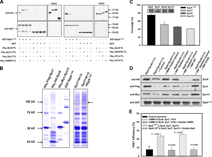
Determination of SipA-mediated formation of functional SNARE complex required for membrane fusion. (A) To identify SipA-binding SNARE partners, GST-SipA1–435 was immobilized on beads, and binding with indicated His6-SNARE proteins was determined as described in Materials and methods. His6-Syn8 was used as a positive control. (B) To determine whether SipA, Syn8, Syn7, and Syn13 form SDS-resistant complexes, GST-SipA1–242, His6-HA-Syn8, His6-Myc-Syn13, and His6-FLAG-Syn7 proteins were purified and analyzed by SDS-PAGE without heat denaturation under nonreducing conditions, shown as Coomassie-stained bands (left). Strongly stained bands (asterisks) correspond with the expected size of the monomer of the respective proteins. Purified His6-HA-Syn8, His6-Myc-Syn13, and His6-FLAG-Syn7 proteins were incubated in the presence and absence of GST-SipA1–242 and analyzed by SDS-PAGE as described in Materials and methods. Arrow indicates a band corresponding with high-MW hybrid complex only when SipA1–242 was present (right). (C) Percent coverage of all four proteins in the indicated hybrid complex as revealed by proteomic analysis. Inset shows the Western blot analysis using specific antibodies. (D) To determine formation of functional SNARE complex by SipA with Syn8, Syn13, and Syn7, we analyzed the disassembly of the SNARE complex by NSF. GST-SipA1–242 was immobilized on beads and incubated with His6-HA-Syn8, His6-Myc-Syn13, and His6-FLAG-Syn7 proteins to form a SNARE complex. Subsequently, disassembly of SNARE complex was determined in the presence and absence of untreated, NEM-treated, or NSF-depleted cytosol by Western blot analysis as described in Materials and methods. All results are representative of three independent experiments. (E) To directly demonstrate fusion between SipA and host Q-SNAREs, we determined the fusion of donor liposome containing SipA1–277 with acceptor liposome containing Syn8, Syn13, and Syn7 by fusion-induced lipid mixing using a standard FRET-based assay as described in Materials and methods. The fusion between donor liposomes containing VAMP8 with acceptor liposomes containing Syn8, Syn7, and Vti1B was used as control. All results are mean ± SD of four independent experiments and are expressed as percent FRET efficiency.

