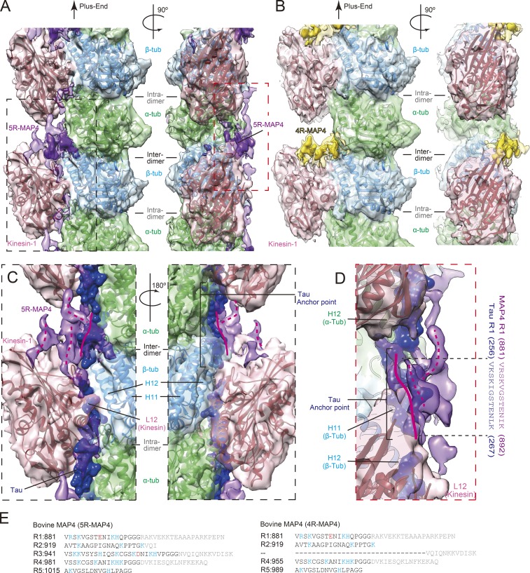
Figure 3.
Structural detail of MAP4–kinesin-1–microtubule binding. (A) Cryo-EM reconstruction of 5R-MAP4–kinesin-1–microtubule complex seen from the right side of the protofilament (left) and from the surface (right). Green, α-tubulin; light blue, β-tubulin; pink, kinesin-1; purple, 5R-MAP4. Higher threshold density for MAP4 is shown in opaque. (B) Cryo-EM reconstruction of 4R-MAP4–kinesin-1–microtubule complex. Green, α-tubulin; light blue, β-tubulin; pink, kinesin-1; yellow, 4R-MAP4. Higher threshold density for MAP4 is shown in opaque. (C) Magnified view of black dotted rectangle in A shown with Tau (blue; PDB ID: 6CVN). A 180°-rotated view is also presented. (D) Magnified view of red dotted rectangle in A shown with Tau. The anchor point sequences of Tau and corresponding sequence of MAP4 are also presented. (E) Amino acid sequences of the tubulin-binding repeat of bovine 5R-MAP4 and 4R-MAP4.

