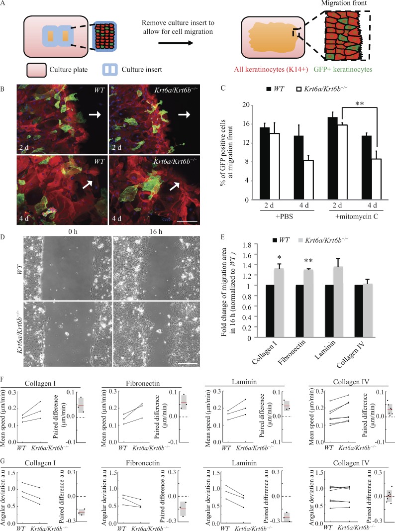
Figure 1.
Increased migration of Krt6a/Krt6b-null keratinocytes is cell autonomous and a result of enhanced speed and more coordinated movement. (A) Schematic of the coculture migration assay. GFP (green) labeled tracer keratinocytes were mixed with either WT or Krt6a/Krt6b-null keratinocytes and plated within a culture insert placed on top of a coverslip coated with type I collagen. K14 (red) labeled all keratinocytes. When cells were confluent, the culture insert was removed to allow for cell migration. The cells at migration front (rectangular area with dashed lines) were imaged, and the percentage of GFP-positive cells within these areas (≥15 images) was quantified. (B) WT and tracer keratinocytes seemed to have equivalent potential for migration, whereas Krt6a/Krt6b-null keratinocytes migrated at a faster rate than tracer keratinocytes. Representative images show cells that were treated with PBS vehicle control. K14 is in red, and DAPI is in blue. GFP (green) staining represents tracer keratinocytes expressing GFP-tagged actin. Arrows point toward the direction of migration. (C) Data represent the mean + SEM for at least three biological replicates. **, P < 0.01, Student’s two-tailed t test. (D) Phase-contrast imaging was performed to monitor cell migration (Video 1). Representative images show WT and Krt6a/Krt6b-null keratinocytes migrating on type I collagen. Bars, 200 µm. (E) Krt6a/Krt6b-null keratinocytes had an increased area of migration when migrating on type I collagen, fibronectin, and laminin but not on type IV collagen. Data represent the mean + SEM of three or more biological replicates. *, P < 0.05; **, P < 0.01, Student’s two-tailed t test. (F and G) PIV analysis showed that compared with a WT cell sheet, a Krt6a/Krt6b-null cell sheet had an enhanced speed of migration on type I collagen, fibronectin, laminin, and to a lesser extent, type IV collagen. Krt6a/Krt6b-null cell sheet was more coordinated than WT cell sheet on type I collagen, fibronectin, and laminin, but not on type IV collagen (Videos 2 and 3). Experiments from the same day were paired and linked with a line. Paired difference was calculated by subtracting value of WT cells from that of Krt6a/Krt6b-null cells.

