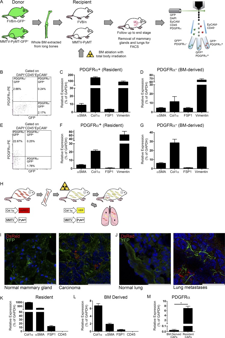
Figure 2.
A subpopulation of CAFs in mammary tumors and lung metastases are BM derived. (A) Scheme of BM transplantation model. 6-wk-old PyMT and FVB/n female mice were transplanted with fresh whole BM isolated from age-matched GFP-PyMT and GFP female mice, respectively. Mice were sacrificed when PyMT recipients had end-stage advanced carcinoma. BM transplantation was repeated five times. (B–G) PDGFRα is a marker of resident fibroblasts. (B) FACS analysis of mammary tumors derived from a BM-transplanted PyMT mouse. n = 4. (C and D) qRT-PCR analysis of fibroblastic markers in the PDGFRα+GFP− (C) and PDGFRα−GFP+ (D) cell populations presented in B. Results show mean ± SD of technical repeats. (E) FACS analysis of metastases-bearing lungs from BM-transplanted PyMT mice. n = 2. (F and G) qRT-PCR analysis of fibroblastic markers in the PDGFRα+GFP− (F) and PDGFRα−GFP+ (G) cell populations presented in E. Error bars represent SD of technical repeats. (H–M) A subpopulation of Col1α+ CAFs in mammary tumors and lung metastases are BM derived. (H) Scheme of BM transplantation model. Following BM ablation with total body irradiation, 6-wk-old PyMT;Col1α-YFP or FVB/n Col1α-YFP female mice were transplanted with fresh whole BM isolated from age-matched PyMT;Col1α-DsRed or FVB/n Col1α-DsRed female mice, respectively. Mice were analyzed when PyMT;Col1α-YFP recipients had advanced carcinoma tumors. BM transplantations were repeated five times (n = 2–4 mice in each cohort). (I) Immunofluorescent staining of resident (YFP) and BM-derived (DsRed) cells in normal mammary glands from FVB/n Col1α-YFP recipients or in mammary tumors from PyMT;Col1α-YFP recipient mice. Bars: 50 µm (left); 25 µm (right). (J) Immunofluorescent staining as in I in normal lungs from FVB/n Col1α-YFP recipients or in lung macrometastases from PyMT;Col1α-YFP recipients. Bars: 50 µm (left); 25 µm (right). For I and J, multiple fields from at least three mice were analyzed. Cell nuclei, DAPI; YFP, Alexa Fluor 488; DsRed, Rhodamine. (K) qRT-PCR analysis of fibroblastic and leukocyte markers in the YFP+ (resident) cells, FACS sorted from mammary tumors in the PyMT;Col1α-YFP recipient mice (n = 2, pooled). Error bars represent SD of technical repeats. (L) qRT-PCR analysis as above in the DsRed+ (BM-derived) cells isolated from recipient mice (n = 2, pooled). Error bars represent SD of technical repeats. (M) qRT-PCR analysis of PDGFRα expression in YFP+ (resident) and DsRed+ (BM-derived) cells isolated from recipient mice (pooled). Error bars represent SD of technical repeats. *, P = 0.05; one-tailed Mann-Whitney test.

