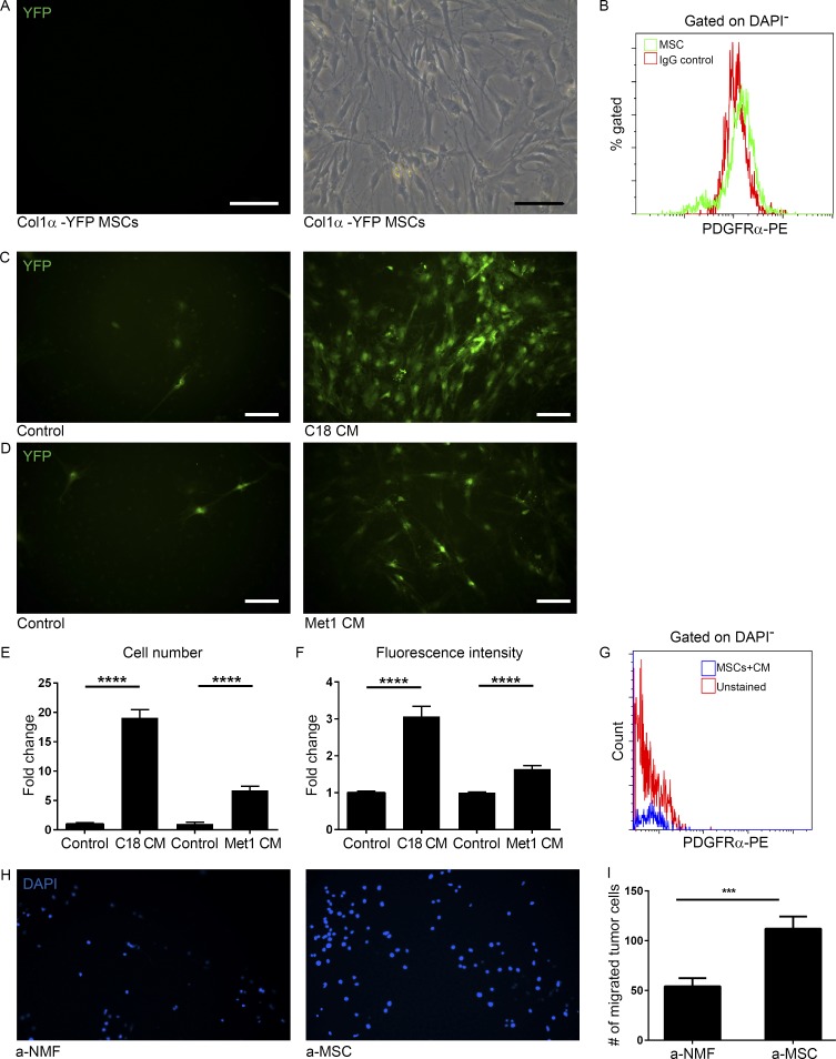
Figure 3.
Tumor cell–secreted factors induce differentiation of BM-derived mesenchymal stem cells to CAFs. (A) Images of cultured mesenchymal stem and progenitor cells (MSCs) produced from total BM of FVB/n Col1α-YFP mice. Light microscopy (right panel) and green fluorescence (left panel). n = 4. Representative of two independent experiments. Bars, 100 µm. (B) FACS analysis of PDGFRα in MSCs. (C and D) Fluorescent microscope images of MSCs that were incubated with C18 CM (C, right) or Met-1 CM (D, right) for 3 wk and compared with controls cultured in 10% FCS medium (left panels). YFP+ cells are shown in green. n = 4. Bars, 100 µm. (E and F) Quantification of YFP+ cell number (E) and fluorescence intensity (F) of images presented in C and D. 30 fields of CM and 10 fields of control were analyzed. Error bars represent SEM. ****, P < 0.0001, two-tailed Mann-Whitney test. (G) FACS analysis of PDGFRα in MSCs incubated with Met-1 CM. (H) Migration transwell assay of Met-1 mammary tumor cells incubated with tumor-activated NMFs (a-NMFs) or MSCs (a-MSCs) for 24 h. Representative images of 24 fields analyzed from duplicate wells. (I) Quantification of data shown in H. Results show mean ± SEM. ***, P = 0.0008; two-tailed Mann-Whitney test.

