Figure 2.

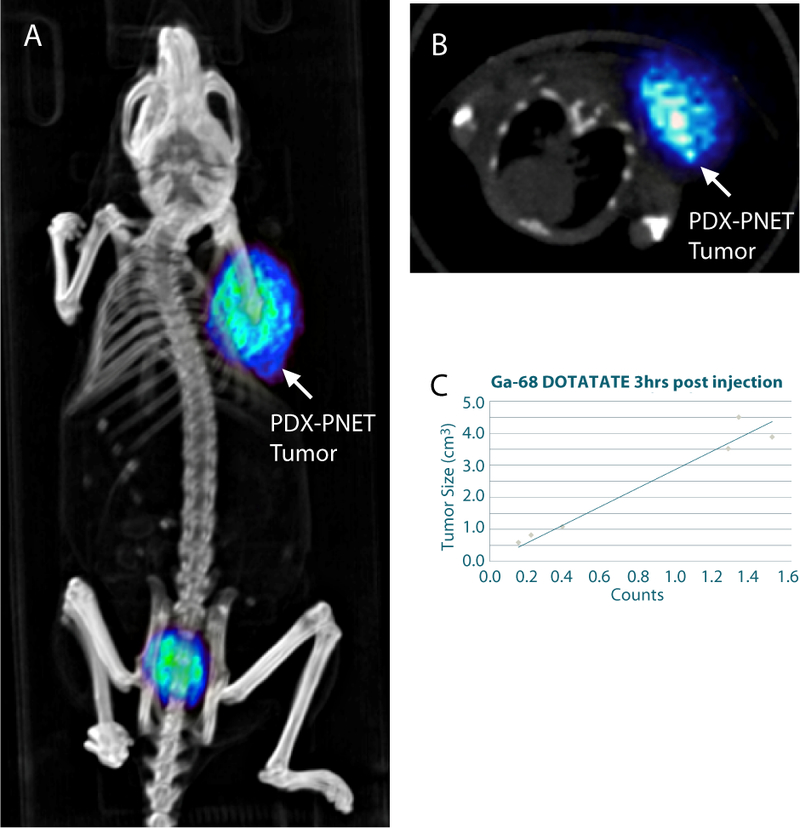
68Ga-DOTATATE PET-CT imaging of the PDX-PNET model. A, 3D rendered PET/CT image showing flank tumor tracer uptake. B, axial view at the level of the tumor. C, relationship between tumor size and normalized 68Ga-DOTATATE PET/CT counts.

68Ga-DOTATATE PET-CT imaging of the PDX-PNET model. A, 3D rendered PET/CT image showing flank tumor tracer uptake. B, axial view at the level of the tumor. C, relationship between tumor size and normalized 68Ga-DOTATATE PET/CT counts.