Figure 1.

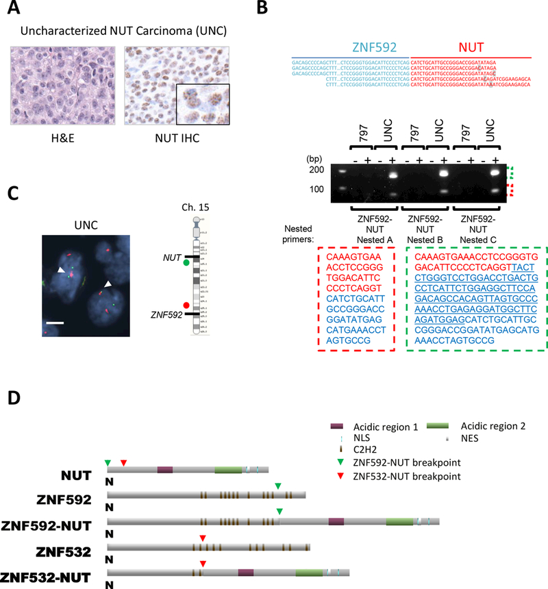
ZNF592-NUT fusion oncogene was discovered in FFPE tumor from a NC patient. A, H&E stained (left) and diagnostic anti-NUT immunohistochemistry (right) of resected NC of the pelvic bone of the 18 year old patient with uncharacterized NUT carcinoma (UNC) (magnification: 400×; inset, 1707×). B, Top: The result of Archer® FusionPlex® analysis indicating the sequence spanning the breakpoint of ZNF592 and NUT. Unique nucleotides in each read were highlighted in grey. Bottom: Gel electrophoresis of nested RT-PCR of TC-797 and UNC with (+) and without (–) reverse transcriptase. Nested PCR using three different nested primer sets (ZNF592-NUT Nested A/ B/ C) on TC-797 and UNC. The sequences corresponding to each band are indicated with red or green dashed lines. ZNF592 and NUT sequences are indicated by red and blue letters, respectively. The underlined sequence was detected only in the larger 200 bp band. C, Dual-color FISH bring-together assay using centromeric 5’ ZNF592 probes (red) and telomeric 3’ NUT (green). White arrows indicate the ZNF592-NUT fusion. magnification: 1000×, scale bar: 5 μm. D, Schematic of the ZNF592-NUT predicted encoded protein in comparison with ZNF532-NUT. Green arrows and red arrows denote ZNF592-NUT breakpoints and ZNF532-NUT breakpoints, respectively.
