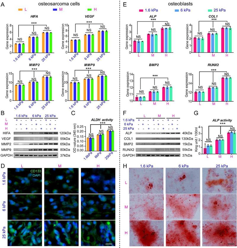
Fig. 4. The tumorigenesis of osteosarcoma cells and osteogenesis of osteoblasts cultured within scaffolds in vitro.

(A) HIF, VEGF, MMP2 and MMP9 mRNA expression in osteosarcoma cells cultured within scaffolds for 7 days. (B) Representative blots of HIFA, VEGF, MMP2, MMP9 and GAPDH in intrascaffold-cultured osteosarcoma cells after 7 days. (C) ALDH activity in intrascaffold-cultured osteosarcoma cells after 7 days. (D) The immunofluorescent staining of CD133 in intrascaffold-cultured osteosarcoma cells after 7 days. Scale bar: 10 µm. (E) ALP, BMP2, COL1 and RUNX2 mRNA expression in osteoblasts cultured within scaffolds for 7 days. (F) Representative blots of ALP, COL1, BMP2, RUNX2 and GAPDH in intrascaffold-cultured osteoblasts after 7 days. (G) ALP activity of intrascaffold-cultured osteoblasts after 7 days. (H) Alizarin red staining of intrascaffold-cultured osteoblasts after 7 days. Scale bar: 10 µm. n=3; mean ± SD; *P<0.05, **P<0.01, ***P<0.001; NS, not significant. L, M and H correspond to low (0.05% GelMA), middle (0.2% GelMA) and high (0.5% GelMA) adhesion ligand density.
