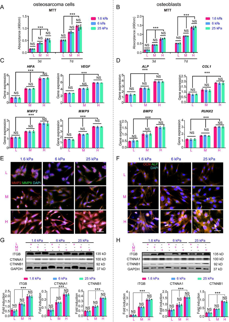
Fig. 8. Behavior of osteosarcoma cells and osteoblasts cultured on the scaffold surfaces.

(A) Cell Proliferation of surface-cultured osteosarcoma cells after 3 and 7 days. (B) Cell proliferation of surface-cultured osteoblasts after 3 and 7 days. (C) HIF, VEGF, MMP2 and MMP9 mRNA expression in osteosarcoma cultured on the surfaces after 7 days. (D) ALP, COL1, BMP2 and RUNX2 mRNA expression in osteoblasts cultured on the surfaces after 7 days. (E) Immunofluorescence staining of MMP2 and MMP9 in intrascaffold-cultured osteosarcoma cells after 7 days. Scale bar: 50 µm. (F) Immunofluorescence staining of ALP and OCN in intrascaffold-cultured osteoblasts after 7 days. Scale bar: 50 µm. (G) Representative blots and fold induction of ITGB, CTNNA1, CTNNB1, FAK and GAPDH in osteosarcoma cells cultured on surfaces after 7 days. (H) Representative blots and fold induction of ITGB, CTNNA1, CTNNB1, FAK and GAPDH in osteoblasts cultured on surfaces after 7 days. n=3; mean ± SD; *P<0.05, **P<0.01, ***P<0.001; NS, not significant. L, M and H correspond to low (0.05% GelMA), middle (0.2% GelMA) and high (0.5% GelMA) adhesion ligand density.
