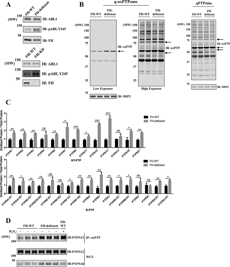
Figure 2.
Multiple classical PTPs are oxidized in FH-deficient PRCC cell lines. A, UOK262 (FH-WT and FH-deficient) cells (upper panels) and YUNK1 (FH-WT and FH-KD) cells (lower panels) were lysed, and the levels of ABL1 and pABL-Y245 were assessed by immunoblotting. B, UOK262 (FH-WT or FH-deficient) cells were processed by q-oxPTPome (left panels) or qPTPome (right panel) and immunoblotted with oxPTP Ab. Arrows indicate potentially highly oxidized proteins in FH-deficient cells. SHP2 serves as a loading control. C, Samples from Fig. 2B were analyzed by MS and label-free quantification. The q-oxPTPome signal was normalized to the qPTPome signal (to adjust for PTP expression). Data represent mean ± SEM (n=3; *p<0.05, **p<0.01, ***p<0.001, ****p<0.0001; unpaired two-tailed t-test). D, UOK262 (FH-WT or FH-deficient) were processed through q-oxPTPome. Samples were immunoprecipitated with oxPTP Ab and immunoblotted for PTPN12. H2O2, 1 mM for 4 min.

