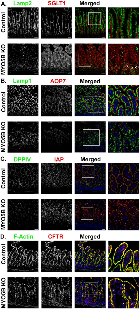
Figure 1: MYO5B KO mice exhibit loss of SGLT1, apical alkaline phosphatase and AQP7 but maintain apical CFTR expression.
(A) Lamp2 positive lysosomes (green) were present below the brush border labeled by SGLT1 (red) in control mice. MYO5B KO mice showed disorganized Lamp2 lysosomes as well as decreased apical SGLT1 and increased intracellular SGLT1. Arrows indicate several SGLT1 positive inclusions surrounded by Lamp2 lysosomes. (B) AQP7, is normally expressed in the brush border of duodenal enterocytes. Apical AQP7 (red) is absent in MYO5B KO mice. Lamp1 lysosomes (green) were aligned below the apical membrane in control mice. MYO5B KO mice had dispersedLamp1 lysosomes. (C) Immunofluorescence staining for IAP (red) in control mice showed clear apical expression. MYO5B KO mice exhibited subapical accumulation of IAP below the apical membrane. DPPIV (green) was present in numerous intracellular inclusions in MYO5B KO mice. (D) CFTR (red) and F-actin (green) are normally expressed in the brush border of enterocytes. Following loss of MYO5B F-actin positive inclusions were observed. CFTR was present in F-actin positive inclusions in MYO5B KO mice. However, CFTR was still present on the apical membrane of MYO5B KO mice. n=4-6 mice per group, scale bars=50 μm.

