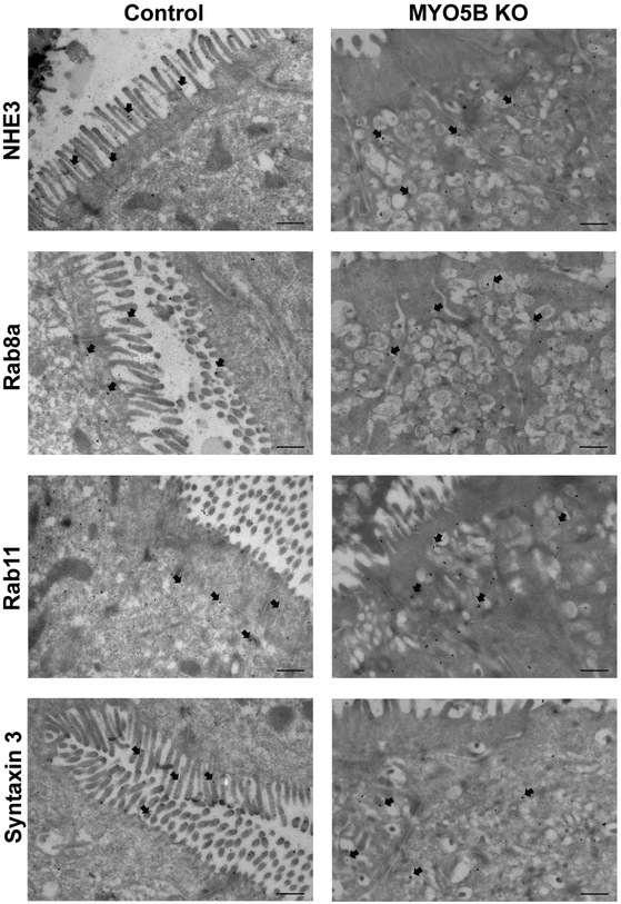
Figure 2: Localization of NHE3, Rab8a, Rab11 and STX3 by immunogold Transmission Electron Microscopy.
NHE3 immunogold labeled microvilli of control mice but was additionally present in subapical vesicles in MYO5B KO mice. Rab8a and Rab11 showed normal subapical distribution in control mice and in MYO5B KO mice a certain accumulation in the aberrant subapical vesicle clusters. STX3 was found on the brush border of control mice but was preferentially located in subapical vesicles in MYO5B KO mice. n=3-4 mice per group, scale bars=500 nm.

