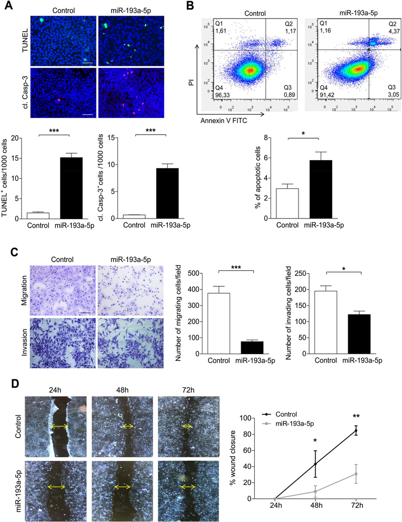
Figure 3.
Overexpression of miR-193a-5p increases apoptosis in vitro. (A) Cell death (TUNEL staining (upper row); cl. Casp-3 staining (lower row)) was analyzed in Huh7 cells transfected with miR-193a-5p and Control siRNA for 72h. (bar = 50 μm). (B) Apoptosis of Huh7 cells transfected with miR-193a-5p was determined by Annexin V/Propidium iodide staining and flow cytometry (n = 3 per group). (C) Transwell migration assay (upper row) or Transwell coated with Matrigel invasion (lower row) assay for Huh7 cells was determined after transfection with miR-193a-5p mimic or Control siRNA for 72 h (n = 3 per group) (bar = 50 μm). (D) Wound-healing assay was performed on Huh7 cells transfected with miR-193a5p mimic and wound closure was monitored over the indicated time post-transfection after treatment with 10 ¼g/ml Mitomycin C for 2 h (n = 3 per group) (bar = 200 μm). Results are represented as mean ± SEM. ns non-significant, * p<0.05, ** p<0.01, *** p<0.001 by 2- tailed, unpaired t test.

