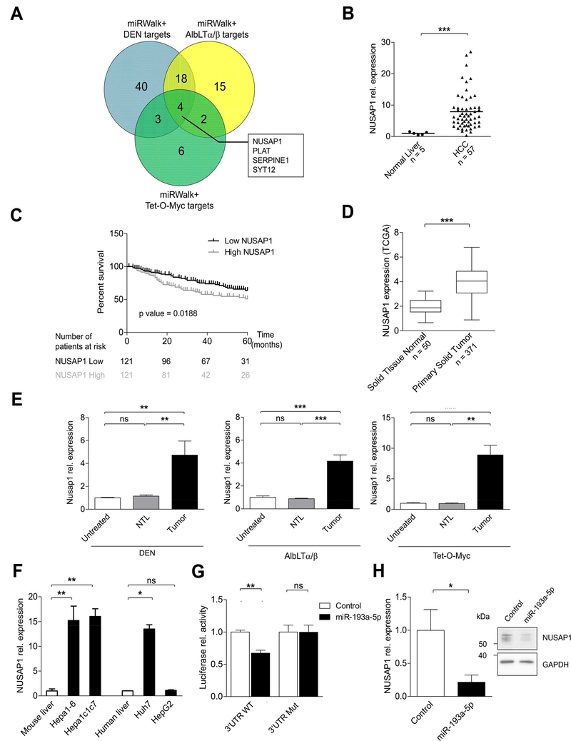
Figure 4.
NUSAP1, a direct target of miR-193a-5p, is upregulated in mouse and human HCC tissues. (A) Venn diagram showing the overlap between predicted targets of miR-193a-5p and genes (threshold: fold change > 2, p value < 0.01) obtained from mRNA-array of DEN, AlbLTα/β and Tet-O-Myc mouse HCC models. (B) Relative expression of NUSAP1 in human HCC (n = 57) compared to normal liver (n = 5) from Affymetrix array. (C) Kaplan-Meier curves for the overall survival of HCC patients (n = 242) plotted against time (months) based on NUSAP1 expression levels. (D) Expression levels of NUSAP1 in 371 HCC patients and solid normal tissue (n = 50) are shown (primary data obtained from TCGALIHC datasets). (E) Relative expression of Nusap1 was measured in non-tumoral liver (NTL) and HCC tumors of mice from DEN, AlbLTα/β and Tet-O-Myc in comparison with untreated mice. (F) Relative expression of NUSAP1 in mouse (Hepa1–6 and Hepa1c1c7) and human (Huh7 and HepG2) HCC cell lines was quantified by qRT-PCR (n = 3 per group). (G) Relative luciferase activity was quantified in Huh7 cells co-transfected with reporter constructs containing either WT or Mutant NUSAP1 together with miR-193a-5p mimic or control (n = 3 per group). (H) Relative expression of NUSAP1 in Huh7 cells transfected with miR-193a-5p mimic was quantified by qRT-PCR and Western Blot (n = 3 per group). Results are represented as mean ± SEM. ns non-significant, * p<0.05, ** p<0.01, *** p<0.001 by 2- tailed, unpaired t test (B, D, G and H); Log-Rank with Mantel-Cox test (C); 1-way ANOVA with Newman-Keuls post-hoc test (E and F).

