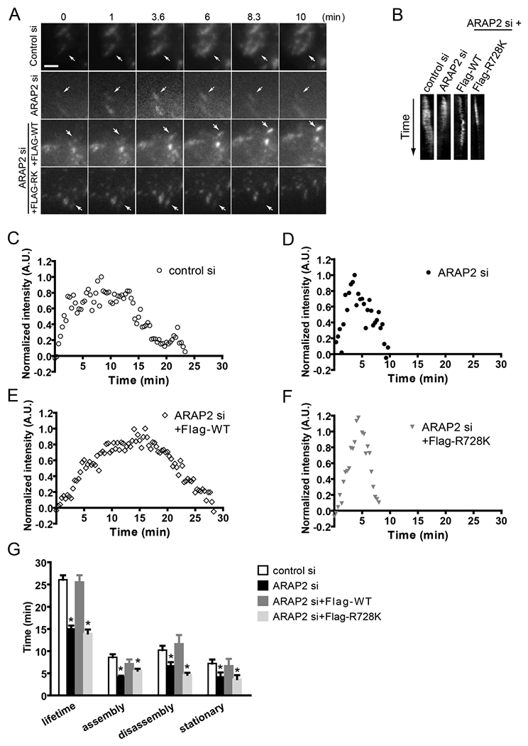
Figure 1. Reduced ARAP2 expression promotes FA turnover.

(A-G) HeLa cells treated with siRNA (control si, ARAP2 si) were transfected with a plasmid for expression of GFP-paxillin and either a vector control or different constructs for the expression of ARAP2 at a ratio of 1:7 (ARAP2 si+Flag-tagged WT, R728K), then plated onto fibronectin-coated (10 μg/ml) coverslips. The next day, images were obtained from a time-lapse TIRF series of GFP-paxillin during FA turnover. Data shown are (A) time-lapse images, (B) kymographs of GFP-paxillin adhesions, (C-F) examples of normalized fluorescent paxillin intensity change over time in the FAs, and (G) the average FA lifetime and length of assembly, disassembly and stationary phase from adhesions in each condition (n=14~19, ± s.e.m.). * p<0.05 indicates significantly different from control siRNA using Student’s t test. Scale bar, 2.5 μm.
