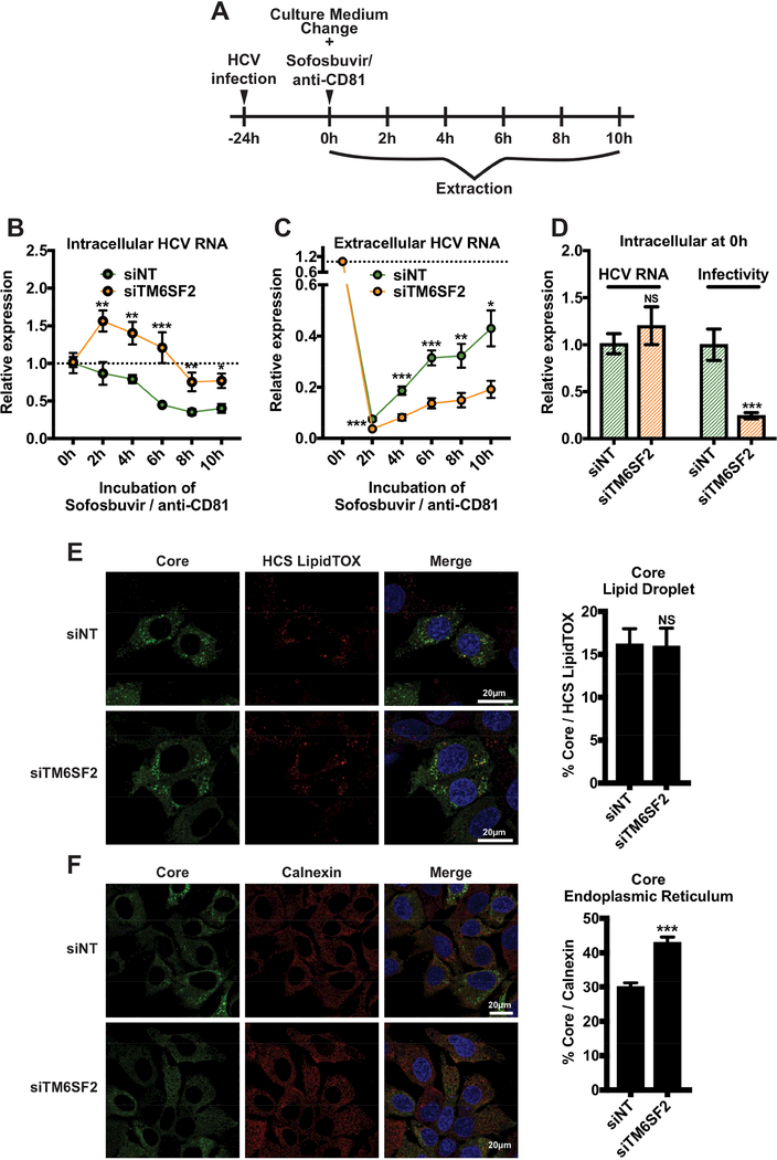
Figure 2: TM6SF2 knockdown blocks the secretion of HCV RNA and infectious particles.
Huh7.5.1 cells were transfected with siRNA as indicated for 72 h and infected with HCV. (A) 24 h post-infection, at time point 0 h, media were changed and supplemented with sofosbuvir and anti-CD81 Ab. Samples were harvested at 2, 4, 6, 8 and 10 h after treatment. (B) Intracellular and (C) extracellular total RNA were extracted and HCV RNA were quantified of by qRTPCR (standardized to 18S RNA). (D) At time point 0 h, the relative expression of HCV RNA was quantified and cell lysates (50 μL) were used to infect naïve Huh7.5.1 cells. At 48 h p.i., total RNAs were then extracted and HCV RNA levels were quantified by qRT-PCR to establish HCV infectivity. HCV RNA and infectivity were compared to the siNT condition. All experiments were independent and performed 3 times in triplicates (n=9). (C) 48 h p.i. cells are fixed, HCV core, LDs (HCS LipidTOX) and ER (anti-calnexin Ab) were stained. Percentages of colocalization between core and LDs or ER were quantified, using Zen software (at least n=64). White bars scale, 20 μm. Shown values are means ± SEM. NS, non-significant; *, P-value < .05; **, P-value < .01; ***, P-value < .001 (Mann Whitney’s test).

