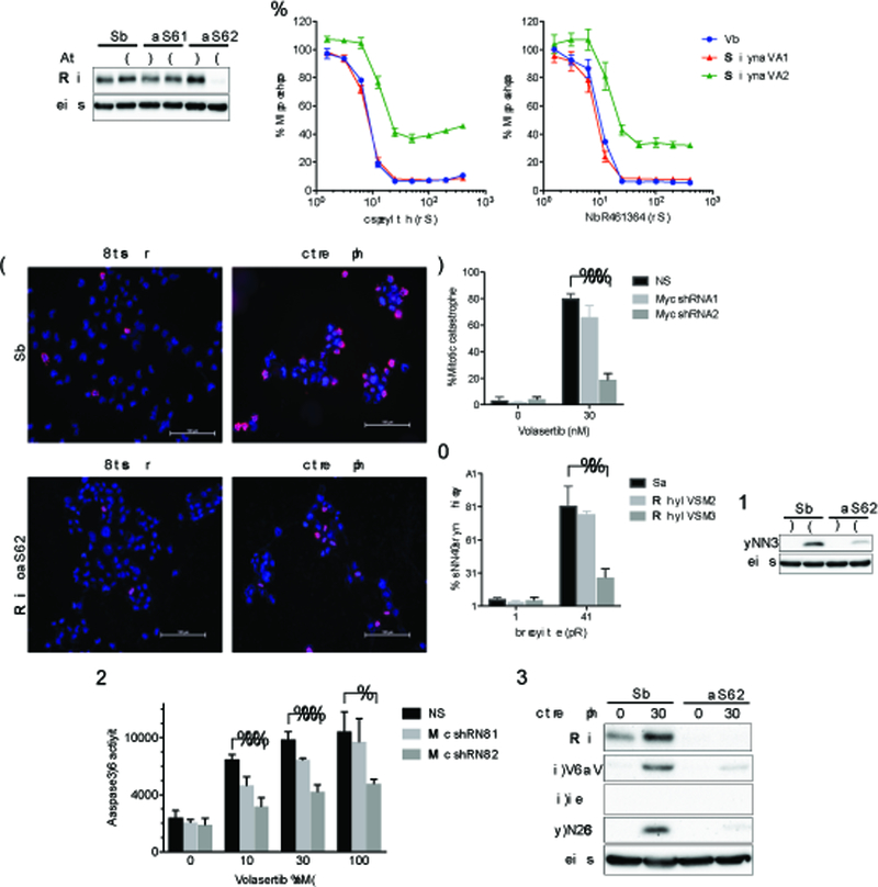
Figure 6. Myc silencing desensitizes glioblastoma cells to PLK1 inhibitors.

(A) Gli36 cells stably transduced with a non-targeting (shNS) or tetracycline-regulatable cMyc shRNA (shRNA1, shRNA2) vectors were established. Cells were lysed after exposed to doxycycline (Dox) for 72 hours. Immunoblotting confirmed a decrease in Myc by shRNA2. Actin was used as a loading control. (B) Gli36-shNS (NS, blue), Gli36-MycshRNA1 (Myc shRNA1, red) and Gli36c-MycshRNA2 (Myc shRNA2, green) cells were treated with different concentrations of Volasertib (left) and GSK461364 (right). Cell viability was evaluated by CellTiter Glo at 72 hours. (C-E) Gli36-NS (NS) and Gli36-MycshRNA2 (Myc shRNA2) cells were treated with/without Volasertib at 30 nM for 24 hours and immunostained for pHH3 (red) and counterstained with DAPI. Scale bars, 100 μm. % cells with abnormal mitosis (mitotic catastrophe) (D) and pHH3 positive cells (E) were evaluated in at least 100 cells per experiment. (F) Immunoblot of pHH3 expression in Gli36-NS and Gli36-MycshRNA2 cells with/without Volasertib treatment (30 nM, 24 hours). Actin was used as a loading control. (G) Gli36NS (NS), Gli36-MycshRNA1 (Myc shRNA1) and Gli36-MycshRNA2 (Myc shRNA2) cells were treated with indicated concentrations of Volasertib for 24 hours. Caspase3/7 activity was evaluated by Caspase-Glo 3/7 assay. (H) Immunoblot of Myc, cleaved PARP (c-PARP), cleaved caspase3 (c-caspase3) and p-H2Ax expression in Gli36-NS (NS) and Gli36-MycshRNA2 (shRNA2) cells after treatment with Volasertib (0, 30 nM) for 24 hours. Actin blot was used as a loading control. In D, E and G, *, P<0.01; **, P<0.005; ***, P<0.001 (student t-test).
