
Fig. 3.
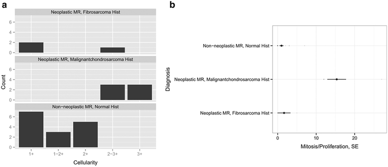
Histopathological classification of neoplastic and non-neoplastic transplants and MRI correlation. Normal and malignant ADSC implants were quantitatively compared based on a cellularity (1+ = paucicellular, 2+ = moderately cellular, 3+ = highly cellular) and b the proliferation rate per high power field, hpf (× 400 magnification). Malignant transplants were defined based on high cellularity and high mitosis rate. Non-neoplastic transplants exhibited low mitosis rates that were the result of cartilaginous proliferation.