Fig. 1.
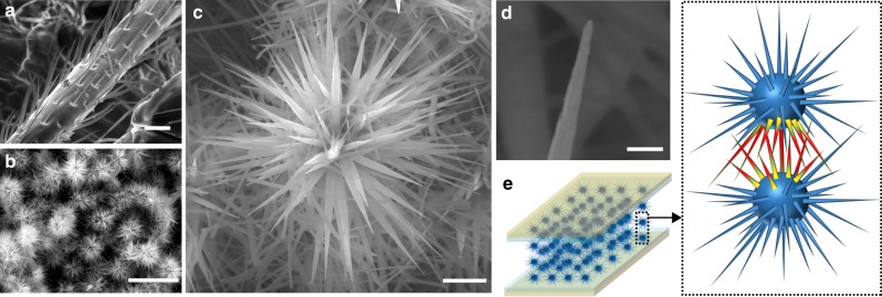
ZnO-microparticle morphology and sensor concept. a Scanning electron microscopy (SEM) image of a leg from an ant, showing the hierarchical distribution of tapering bristles. Scale bar, 20 µm. b SEM image of SUSMs packing together in a thin film. Scar bar, 10 µm. c High-magnification SEM image of one SUSM, showing a forest of nanostructured spines. Scale bar, 1 µm. d Zoom-in SEM image of the tip of a spine. Scale bar, 100 nm. e (Left panel) Schematic of a sensor device made from a SUSM thin film sandwiched between two electrodes. (Right panel) Schematic highlights the resistance modulation at local spine–spine sites induced by mechanical stimuli, with the color gradient indicating induced strain

