Figure 2.
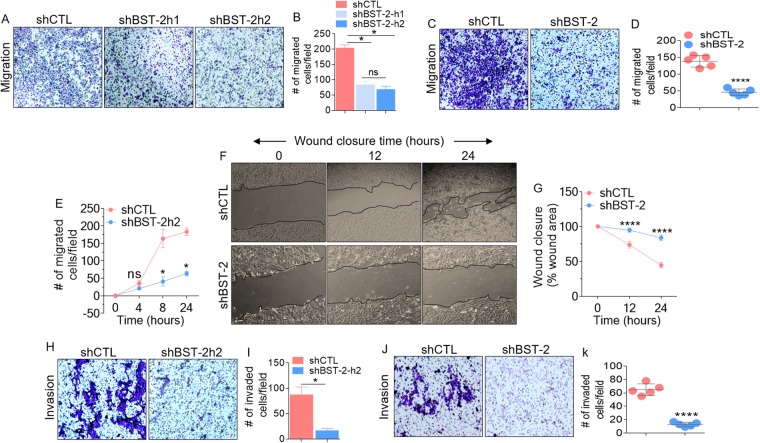
BST-2 expression promotes proliferation-independent migration and invasion of breast cancer cells: (A) Giemsa-stained images of migrated MDA-MB-231 cells stably expressing scramble shRNA control (shCTL) and two different BST-2-targetting shRNA sequences (shBST-2h1 and shBST-2h2). (B) Quantification of migrated cells in panel A. (C) Microscopic images of Giemsa-stained 4T1 cells and (D) Graphs of 4T1 migration events in panel C. (E) Time-dependent trans-well migration of MDA-MB-231 cells. (F) Images showing kinetics of 4T1 wound closure. Black lines on images depict wound border (0 h) and extent of wound closure (12 h–24 h). (G) Quantitative depiction of wound closure events in panel F. (H,I) Images and quantitation of MDA-MB-231 invasion events. (J,K) Representative images and quantitation of 4T1 invasion. Representative images were taken at 4x magnification. Images used for Image J quantitation of migration and invasion were taken at 20x. Events from five different fields were analyzed. Error bars represent standard deviations. Significance was taken at P < 0.05* and P < 0.0001****. Experiments were repeated more than three times with similar results.

