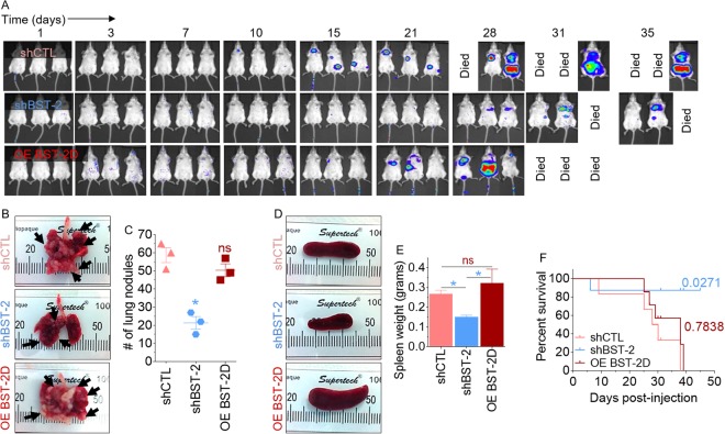
Figure 7.
BST-2 promotes metastasis independent of primary tumor: (A) Representative image of metastases detected after injection of 5-week old BALB/c mice with 300,000 of 4T1 cells expressing shCTL, shBST-2 or OE BST-2D via tail vein. n = 3 for each condition. Tumor cells tracked in vivo with IVIS imaging at different time points. (B) Representative gross images of lungs showing visible pulmonary nodules (arrows). (C) Quantification of lung colonization events in mice described in panel B. (D,E) Gross images and weight of spleens of mice described in panel A. (F) Kaplan-Meier survival plot of mice described in panel A. Numbers are P values relative to shCTL group. Error bars represent SEM and significance was taken at P < 0.05*. ns = not significant.

