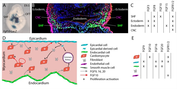
FIGURE 1.
FGF10 signaling in the developing heart. (A) Lateral whole-mount view and (B) transverse section of an embryo carrying an Fgf10-LacZ transgene (Kelly et al., 2001) at embryonic day E9.5. Fgf10 transgene expression is observed in second heart field (SHF) progenitor cells, which are located in pharyngeal mesoderm adjacent to pharyngeal endoderm, and in the outflow tract (OFT). (B) Immunofluorescence on transverse section of an E9.5 embryo carrying an Fgf10-LacZ transgene, at the level of the dotted line in (A). The anti-AP-2α (pink) antibody was used to detect cardiac neural crest (CNC) cells and ectodermal cells and the anti-β galactosidase (green) antibody to visualize SHF cells. (C) Table showing the overlapping expression patterns of key FGF ligands and receptors at E9.5 in the SHF and surrounding tissues. (D) FGF signaling role in fetal heart development. (E) Table showing the overlapping expression patterns of key FGF ligands and receptors in the fetal heart.

