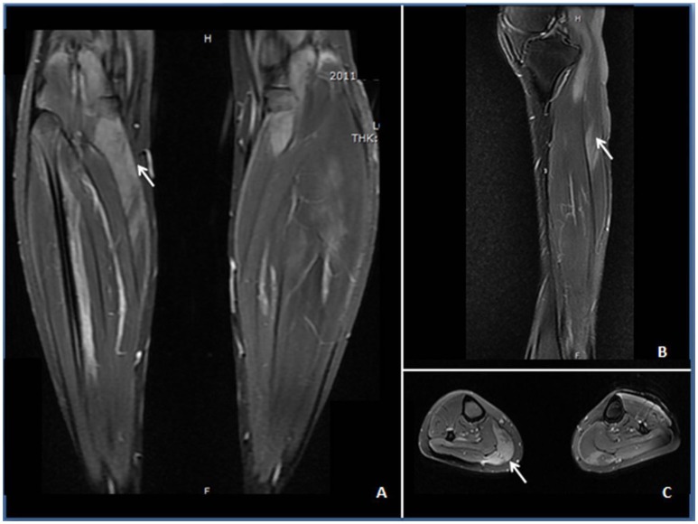
Figure 1.
STIR (A) and T2-weighted images (B,C) demonstrating edema (↑) in the anterior and posterior calves of the patient. (A) Increased STIR image signaling in the gastrocnemius with unsymmetrical involvement. (B) Increased intramuscular T2 image signaling within the anterior tibial muscle at the sagittal section. (C) Patchy T2-weighted hyperintense area in the gastrocnemius, soleus, and anterior tibial muscles.

