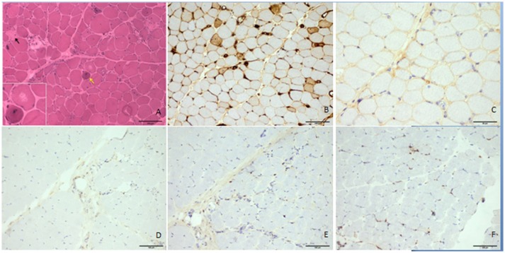
Figure 2.
Histologic features of the patient's quadriceps femoris. The hematoxylin and eosin–stained frozen section demonstrated necrotic fibers (▴), regenerating myofibers (↑), atrophic myofibers, and unusual vacuoles in some degenerated myofibers without prominent lymphocytic infiltrates (A). MHC I was upregulated in some myofibers (B), but there was no prominent accumulation of C5b9 in the myofibers (C). CD4, CD8, and CD68 antibodies did not obviously positively stain the infiltrates (D–F, respectively). Scale bars: 100 μm (A–F) and 50 μm (C).

