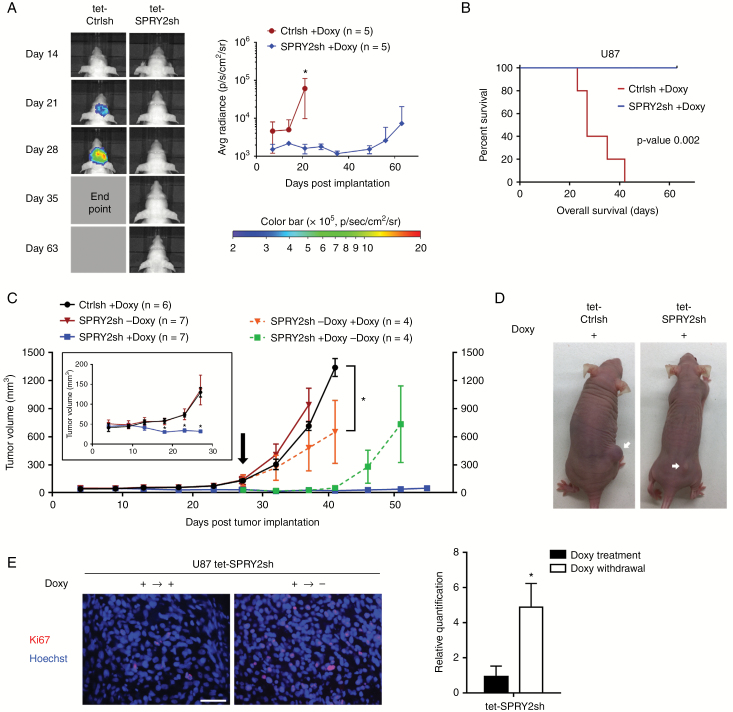
Fig. 5.
Downregulation of SPRY2 inhibits U87 intracranial and subcutaneous tumor growth. (A) U87 tet-Ctrlsh or tet-SPRY2sh cells (0.5 million) expressing RNAi-resistant luciferase were intracranially injected in nude mice (n = 5/group) with administration of doxycycline. Representative image of bioluminescence at the indicated time point (left panel). Quantification of signal intensity presented as photons/sec/cm2/surface radiance, *P < 0.05, mean ± SD (right panel). (B) Kaplan‒Meier survival curve; **P < 0.01 by the log-rank test. (C) U87 tet-Ctrlsh or tet-SPRY2sh cells (2 million) were subcutaneously injected in nude mice (n = 6–7/group) with or without administration of doxycycline. Early stage of subcutaneous tumor development is displayed (inset). Later tumor development following doxycycline treatment in 4 out of 7 previously doxy-untreated tet-SPRY2sh tumor-bearing mice is highlighted in orange. Tumor growth after doxycycline withdrawal in 4 out of 7 doxy-treated tet-SPRY2sh tumor-bearing mice is highlighted in green. *P < 0.05, mean ± SD. (D) Representative pictures of tet-Ctrlsh or tet-SPRY2sh tumor-bearing mice (+Doxy) are shown (arrows indicate tumor). Scale bar = 50 μm. (E) Representative images and quantification of Ki67-positive cells in U87 tet-SPRY2sh tissues (± Doxy). *P < 0.05.

