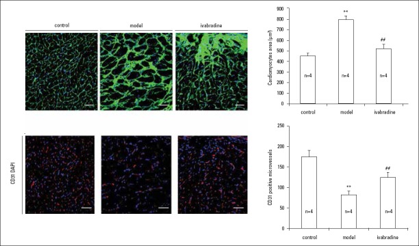
Figure 3.
Effects of IVA on cardiomyocyte hypertrophy and capillary density in MI mice.
(a) Wheat germ agglutinin staining was used to display the structure and size of cardiomyocytes. Sections were from papillary muscle. Scale bar: 40 µm; (b) Quantitation of cross areas of cardiomyocytes (n=4); (c) CD31 staining was used to show capillary density. Scale bar: 40 µm; (d) Quantitation of capillary density (n=4). Data are given as means±SEM. **P<0.01 vs. control group, ##P<0.01 vs. model group

