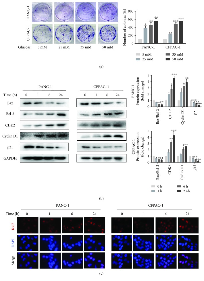
Figure 1.
High glucose promotes pancreatic cancer cell proliferation. (a) Cell viability was measured by colony formation assay after exposure to various concentrations of glucose that range from 5 mM to 50 mM. (b) Western blotting results of Bax, Bcl-2, CDK2, cyclin D1, and p21 in PANC-1 and CFPAC-1 cells after incubating in HG media for 0, 1, 6, or 24 h. (c) The fluorescence intensity of Ki67 (red) after culturing with HG media for 0, 1, 6, and 24 h was detected using immunofluorescence microscopy (×400). Cell nuclei were counterstained with DAPI (blue). Data are presented as mean ± standard error of the mean (SEM) (n = 3). ∗p < 0.05, ∗∗p < 0.01, and ∗∗∗p < 0.001, compared with the 0 h group.

