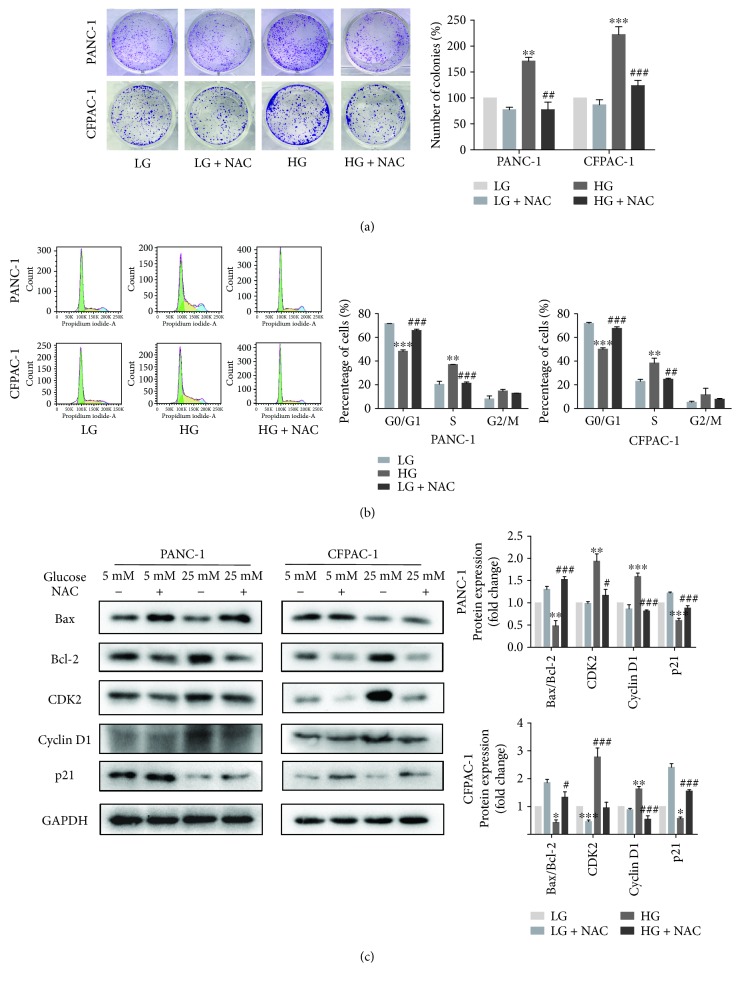
Figure 3.
Suppression of ROS activation would reduce cell proliferation. (a) Colony formation assay following PANC-1 and CFPAC-1 cells incubated in LG and HG media in the presence or absence of 5 mM NAC. (b) Cell cycle distribution was performed by flow cytometric analysis. After stimulating with high glucose and NAC for 24 h, cells were collected and fixed with 70% ethanol for PI staining. The cell analysis was carried out using flow cytometry, and the results are shown as the percentage of cells in each phase of the cell cycle. (c) Cells were treated as described in (a), and then the proteins of Bax, Bcl-2, CDK2, cyclin D1, and p21 were detected by Western blotting. Data are presented as mean ± SEM (n = 3). ∗p < 0.05, ∗∗p < 0.01, and ∗∗∗p < 0.001, compared with the LG group. #p < 0.05, ##p < 0.01, and ###p < 0.001, compared with the HG group.

