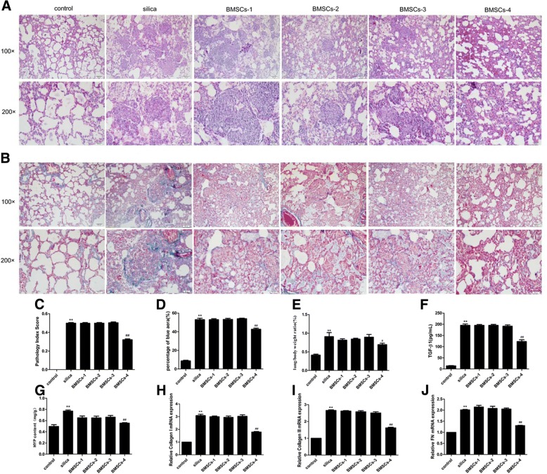
Fig. 3.
Optimal dose and timing of BMSCs administration to attenuate silica-induced pulmonary fibrosis. H&E staining (a) and Masson’s trichrome staining (b) in the rat lungs at 15 days after silica instillation. Light micrograph magnification × 100 and × 200. Scale bar, 100 μm and 50 μm. Quantitatively analyzed image stained in H&E (c) or Masson’s trichrome (d). The pathology index of lung tissue and the fibrotic areas were significantly increased in the silica group compared with the control group, but decreased in the BMSCs-4 group. The ratio of lung/body weight (e), level of TGF-β1 (f), content of HYP (g), expression of collagen I (h), collagen III (i), and FN (j) were significantly increased in the silica group in comparison with the control group, but were decreased in the BMSCs-4 group. BMSCs (2 × 10⁶ cells, days 1 and 4) attenuated silica-induced pulmonary fibrosis. Values are expressed as mean ± SD, n = 8. **p < 0.01 compared with the control group; #p < 0.05, ##p < 0.01 compared with the silica group

