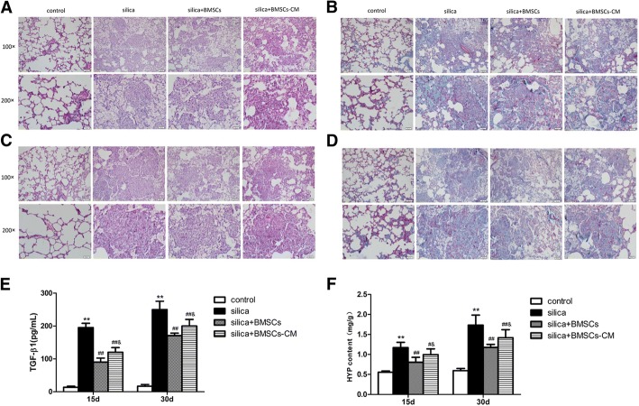
Fig. 5.
BMSCs-CM inhibited silica-induced pulmonary fibrosis in rats. a, c H&E staining, (a) 15 days after instillation; (c) 30 days after instillation. b, d Masson’s trichrome staining. The result revealed that the collagen deposition (blue areas) was significantly increased in the silica group but decrease in the silica + BMSCs-CM group on 15 (b) and 30 days (d). Light micrograph magnification × 100 and × 200. Scale bar, 100 μm and 50 μm. e, f The levels of TGF-β1(e) and content of HYP (f) were increased in the silica group compared with those in the control group, but they were decreased after the addition of BMSCs-CM (p < 0.01). The data were presented as the means ± SD. **p < 0.01 versus the control group; ##p < 0.01 versus the silica group; &p < 0.05 versus the silica + BMSCs group

