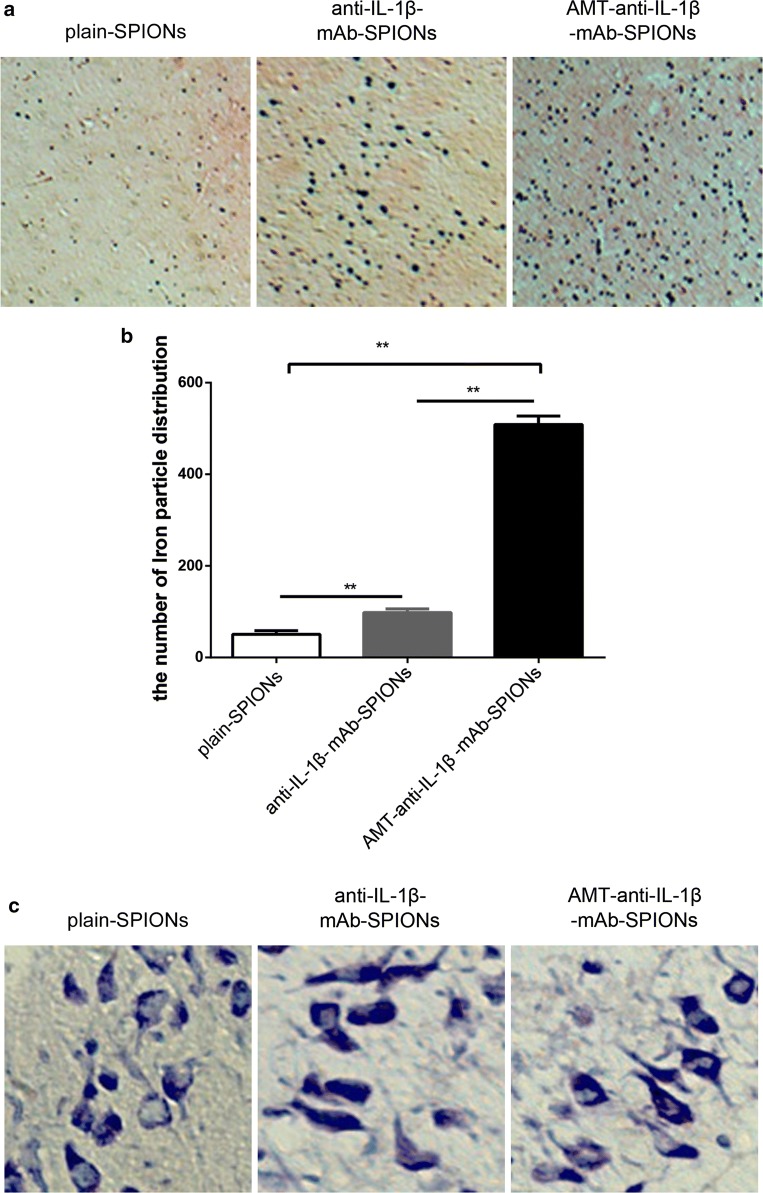
Fig. 2.
Perl’s iron staining to evaluate the iron particles and the corresponding statistical drawing. The iron particles were most densely distributed in the AMT-anti-IL-1β-mAb-SPION group (a), and the statistical analysis was consistent with this finding. b *p < 0.05, **p < 0.01, magnification ×40. Nissl staining to evaluate neuronal morphology and loss. These phenomena of Nissl bodies shrank, and the abnormal neuronal morphology was improved but not very obvious after injection of AMT-anti-IL-1β-mAb-SPIONs than after injections of anti-IL-1β-mAb-SPIONs and plain-SPIONs (c). Magnification ×400

