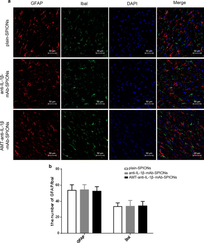
Fig. 3.
Immunofluorescence staining of GFAP and Ibal and the corresponding statistical drawing. Astrogliosis and microglial activation involving cell hypertrophy and cell proliferation were assessed in three groups (a), and there was no significant difference among the groups (p > 0.05, b). Magnification ×200

