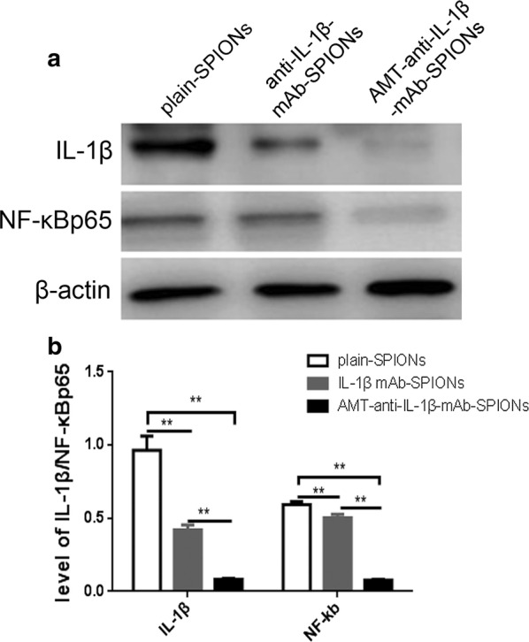
Fig. 5.

Western blot exposure imaging and the corresponding statistical drawing. We normalized IL-1β and NF-ĸB p65 to the corresponding amounts of β-actin. The IL-1β and NF-ĸB p65 concentrations were more clearly decreased in the AMT-anti-IL-1β-mAb-SPION group than those in the anti-IL-1β-mAb-SPION and plain-SPION groups (**p < 0.01, a, b)
